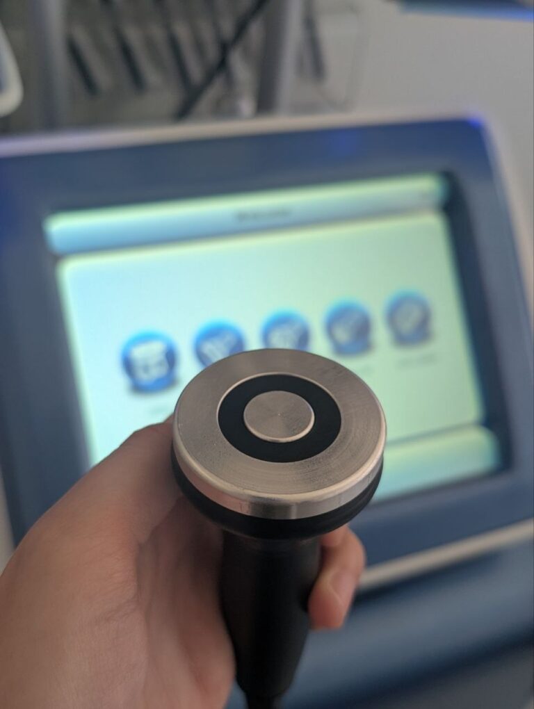
There’s a quiet confidence that comes with wearing a skirt or shorts without a second thought. But for so many of my patients, tiny visible veins on the legs or face chip away at that confidence.

These little vessels—called varicosities, spider veins, or telangiectasias—are incredibly common, and they’re rarely a serious medical concern. Yet they bother people deeply, and I understand why.
In the aesthetic clinic where I’m currently working, one of the most satisfying transformations I witness isn’t a dramatic facelift or a complete hair removal journey; it’s the gentle, precise erasure of these tiny vascular threads that have been quietly stealing someone’s comfort in their own skin.
The technology I trust most for this task is the Nd:YAG laser. That same 1064 nm wavelength we explored in hair removal for dark skin now takes center stage in a completely different role—as a master of vascular precision.
But before we dive into how the laser works, I want to address the question that sits silently in every consultation room: are these veins just a cosmetic problem, or do they mean something is wrong?
Beyond Aesthetics: When Varicosities Signal Something Deeper
This is the question I hear most often, usually phrased as a half-apology: “I know it’s just cosmetic, but they really bother me.”
My response is always the same: let’s talk about that. Because sometimes, these visible vessels are indeed nothing more than a cosmetic concern. And sometimes, they are the first visible whisper of an underlying condition that deserves medical attention.
Simple Telangiectasias (Spider Veins):
These are the fine, red, blue, or purple thread-like veins that sit flush with the skin or bulge only slightly. They are typically less than 1 millimeter in diameter and are most often found on the thighs, calves, ankles, and face.
In the vast majority of cases, these are benign and asymptomatic.
They do not cause pain, they do not indicate deep vein disease, and they do not progress to serious medical conditions. They are, essentially, a cosmetic finding.
However, even here, there is nuance. A sudden shower of new spider veins on the medial ankle, especially when accompanied by itching or a dull ache, can sometimes be an early warning sign of venous insufficiency deeper in the leg, and warrants a Doppler ultrasound evaluation.
Reticular Veins (Feeder Veins):
These are slightly larger, bluish or greenish veins, 1 to 3 millimeters in diameter, that often sit deeper in the skin and can be seen feeding clusters of spider veins. They do not protrude above the skin surface.
On their own, reticular veins are also usually asymptomatic and considered cosmetic. But when they become tender to the touch or are associated with localized discomfort after prolonged standing, they can represent the very beginning of superficial venous incompetence.
Varicose Veins (True Varicosities):
This is where the line between cosmetic and medical can blur definitively. Varicose veins are dilated, tortuous veins greater than 3 millimeters in diameter that bulge above the skin surface.
They are not simply a cosmetic issue; they are a manifestation of underlying venous reflux disease. The one-way valves inside the veins have failed, allowing blood to pool and venous pressure to rise.
Clinical symptoms can include:
Aching, heavy legs that worsen at the end of the day
Throbbing, burning, or cramping sensations
Itching around the affected veins
Restless legs, particularly at night
Swelling of the ankles and feet
Skin changes such as hyperpigmentation (hemosiderin deposition), venous eczema, or lipodermatosclerosis (hardening and darkening of the lower leg skin)
When to Seek Medical Evaluation:
A 2018 clinical review in Circulation emphasized that untreated chronic venous insufficiency can progress to venous leg ulcers, superficial thrombophlebitis (painful clot formation in a surface vein), and, rarely, deep vein thrombosis.
If your leg veins are causing pain, heaviness, swelling, or skin changes, or if you have a family history of venous disease or blood clots, you should be evaluated with a venous duplex ultrasound by a vascular specialist before considering any laser treatment.
Treating the surface veins without addressing underlying reflux is like painting over a water leak—the aesthetic result will be short-lived, and the underlying condition may worsen silently.
The Reassuring Truth: For the majority of patients who walk into my clinic, their visible veins are indeed benign and asymptomatic. The Nd:YAG laser is a beautiful tool for precisely these cases. But my responsibility as a doctor is to ensure we are not beautifying the surface while ignoring the depths. That’s why every consultation begins with a clinical history and a visual assessment. If there is any suspicion of deeper venous disease, a referral is made before a laser is ever turned on.
The Physics of Precision: Why 1064 nm is Uniquely Suited for Vessels
You might remember from our hair removal guide that the Nd:YAG laser operates at a wavelength of 1064 nanometers.
In that context, we celebrated it for safely bypassing epidermal melanin to reach deep hair follicles in dark skin.
Now, we celebrate it for a different, equally elegant reason: its unique relationship with hemoglobin.
A Secondary Target, A Primary Result:
At 1064 nm, melanin absorption is low, which means the laser beam passes gently through the skin’s surface without burning it.
But deeper down, the wavelength is selectively absorbed by its primary target in vascular treatments: oxyhemoglobin, the oxygen-carrying protein inside your red blood cells.
This absorption creates a controlled thermal injury inside the unwanted vessel, causing it to coagulate, collapse, and eventually be harmlessly cleared away by the body’s natural cleanup systems over several weeks.
Deep Penetration: The 1064 nm wavelength penetrates deeply into the dermis, reaching depths of 4 to 6 millimeters.
This is crucial because many leg veins, particularly the reticular “feeder” veins that supply spider veins, sit deeper than shorter wavelengths can effectively reach.
Alexandrite (755 nm) or KTP (532 nm) lasers simply cannot reach these deeper vessels with enough power.
The Evidence: A 2021 study published in Lasers in Surgery and Medicine investigated the histology of 1064 nm laser on leg telangiectasias.
Biopsies taken immediately after treatment showed selective coagulation and destruction of the target vessels while the surrounding dermal tissue and overlying epidermis remained entirely intact.
This microscopic precision is the definition of a safe, targeted therapy.
Treating Leg Varicosities and Spider Veins: A Proven Track Record
When it comes to the lower limbs, Nd:YAG is the workhorse. Leg veins can be notoriously stubborn because they are often under higher venous pressure and can vary wildly in diameter, from tiny red telangiectasias less than 0.5 mm to larger blue reticular veins up to 4 mm.
The Clinical Reality: Sclerotherapy remains a gold-standard first-line treatment for many leg veins, but the Nd:YAG laser shines where needles cannot safely go.
It’s my preferred tool for very fine, needle-shy spider veins (matting), for veins located on the ankles or feet where sclerotherapy carries a higher risk of ulceration, and for patients who have failed or are allergic to sclerosing agents.
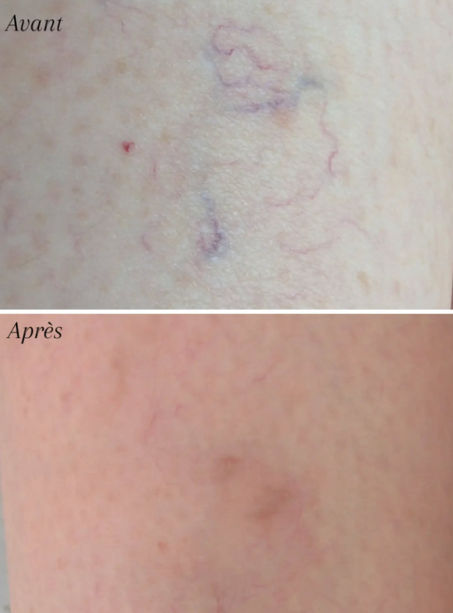
The Evidence: A comprehensive 2020 clinical study in the Journal of the European Academy of Dermatology and Venereology evaluated the long-pulsed 1064 nm Nd:YAG laser on 150 patients with lower extremity spider veins.
After three treatment sessions, independent physician review noted greater than 75% clearance in 82% of treated areas, with results maintained at a 12-month follow-up.
Patients reported high satisfaction, and side effects were limited to transient erythema and mild purpura in a small percentage of cases.
What You’ll See and Feel:
During treatment, you’ll feel a quick snapping sensation with each pulse, accompanied by immediate visible blanching or darkening of the vessel—a positive sign that the blood inside has coagulated.
Over the next 4 to 8 weeks, the treated vessel fades from a dark line to a faint shadow, and then disappears entirely as your body’s lymphatic system resorbs it. Some larger vessels may require two or three sessions for full clearance.
Facial Telangiectasias and Cherry Angiomas: Gentle Enough for Delicate Skin
While leg veins demand deep penetration, facial vessels require a different kind of mastery: gentleness.
The skin on the face is thinner, more sensitive, and more visible. The Nd:YAG laser, with its long wavelengths and adjustable pulse durations, can be calibrated to treat facial vascular lesions without the bruising or downtime that other technologies can cause.
Facial Telangiectasias: These tiny broken capillaries, often found around the nostrils and cheeks, are exquisitely responsive to Nd:YAG.
A 2022 study in Dermatologic Surgery demonstrated that a single pass of a long-pulsed 1064 nm laser with a small spot size and appropriate fluence resulted in immediate vessel disappearance in over 90% of facial telangiectasias, with minimal discomfort and no scarring.
Cherry Angiomas (Campbell de Morgan Spots): Those small, bright red, dome-shaped papules that appear on the trunk, arms, and face with age are an absolute pleasure to treat with Nd:YAG.
A 2024 retrospective review in the Journal of Cosmetic Dermatology confirmed that the long-pulsed 1064 nm laser is a safe and effective modality for cherry angiomas, achieving complete clearance in a single session for lesions smaller than 2 mm, with no post-treatment textural changes to the skin.
Why I Love This Application: For a patient who has been self-conscious about a single prominent red dot on the tip of their nose or a cluster of tiny vessels on the cheek, a single minute of precise laser work can be genuinely life-changing.
It’s a small act of aesthetic medicine with a disproportionately large emotional payoff.
Safety, Downtime, and the Importance of a Trained Hand
Skin Cooling is Non-Negotiable: The 1064 nm beam does generate heat, and protecting the epidermis is paramount. Modern Nd:YAG devices incorporate continuous contact cooling, cryogen spray cooling, or chilled gel to shield the skin surface. This is not a luxury; it’s a safety requirement.
Understanding Post-Treatment Changes: Immediately after treatment, you can expect redness and swelling that typically resolves within 24 to 48 hours.
Purpura (tiny bruises) can occur, especially when treating larger leg vessels with high fluences. For some leg vein protocols, a degree of purpura is expected and correlates with successful vessel destruction, but this should be discussed clearly before treatment. Using a longer pulse duration can minimize bruising, and for facial vessels, I often adjust parameters to achieve clearance with minimal to no purpura, allowing my patients to return to social life the same day.
The Contraindications: Nd:YAG is not suitable for everyone. Patients with active skin infections, a history of keloid scarring in the treatment area, pregnancy, or photosensitivity disorders should not be treated.
A thorough medical history and skin assessment before treatment is not optional—it’s a standard of care.